Introduction
A nanoparticle formulation of docetaxel (DTX) was designed in our lab to address the strengths and limitations of current taxane delivery systems: PEGylation, high drug conjugation efficiency (>30 wt %), a slow-release mechanism, and a well-defined and stable nanoparticle identity were identified as critical design parameters. The polymer conjugate was synthesized with carboxymethylcellulose (CMC), an established pharmaceutical excipient characterized by a high density of carboxylate groups permitting increased conjugation of a drug. CMC was chemically modified through acetylation to eliminate its gelling properties and to improve solvent solubility, enabling high yield and reproducible conjugation of DTX and poly(ethylene glycol). (Ernsting et al., 2011; http://pubs.acs.org/doi/abs/10.1021/bc200284b)
Some existing limitations with the conjugation with polysaccharides include low drug loading (<20%), biologically active polymer chemistry (i.e., heparin and hyaluronan), or rapid release (>20–30% per day). Carboxymethylcellulose (CMC) is an attractive candidate polymer to address the existing limitations as a conjugate backbone, as CMC is approved for use in parenteral formulations, is not biologically active like heparin or hyaluronic acid, and has a high degree of substitution of carboxylic acids for drug conjugation, enabling the incorporation of a high drug content. (Ernsting et al., 2012; http://www.sciencedirect.com/science/article/pii/S0142961211012774)
Detailed Chemistry and Mechanism of Action of Cellax
As previously discussed, Cellax self-assembles into 120nm particles. Cellax is able to adsorb onto albumin and can do so much more efficiently than free DTX (4-fold increased binding). Cellax uptake is mediated through albumin and secreted protein acidic and rich in cysteine (SPARC), an albumin ligand). In the absence of SPARC, Cellax uptake was independent of albumin. However, in the presence of SPARC, a greater increase in Cellax uptake was observed if albumin was present. The internalization mechanism occurs through a clathrin-mediated endocytotic mechanism into endo-lysosomal and autophagosomal compartments. Further experiments showed that tumor growth inhibition was correlated to levels of SPARC in the tumor (R2 > 0.9). (Hoang et al., 2015; http://www.sciencedirect.com/science/article/pii/S0142961215003877)
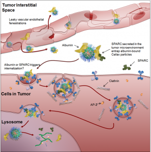
Fig 1. Proposed mechanism of delivery for Cellax nanoparticles. Cellax adsorbs albumin in circulation and is deposited within the tumor interstitium via exploitation of the enhanced permeability and retention (EPR) effect. SPARC produced in the tumor microenvironment binds to the surface albumin on the Cellax nanoparticles and thus traps the particles in the tumor. Cellax is internalized via a clathrin-mediated mechanism and finally ends up in the endolysosomal compartment, where the polymer is broken down and the drug is released.Image from Hoang et al. Biomaterials 2015;59:66-76. doi: 10.1016/j.biomaterials.2015.04.032.)
Pharmacokinetics and Biodistribution of Cellax
Cellax displayed favorable pharmacokinetic and biodistribution properties compared to free docetaxel and Nab-PTX (a nanoparticle formulation of albumin-bound paclitaxel already approved for clinical use). Cellax displayed higher area-under-curve and lower clearance as well as improved uptake into desired target tumors. (Yang et al., 2017: https://www.ncbi.nlm.nih.gov/pubmed/27873118)
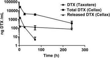
Fig 2: Pharmacokinetic comparison of free docetaxel compared to Cellax-DTX. Cellax-DTX displayed prolonged pharmacokinetics after 40mg DTX/kg infusion of both formulations. From Ernsting et al. Biomaterials 2012;33(5):1445-54.
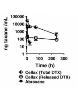
Fig 3: Pharmacokinetic comparison of Abraxane (nab-PTX) compared to Cellax-DTX. Cellax-DTX displayed prolonged pharmacokinetics after 40mg taxane/kg infusion of both formulations. From Ernsting et al. JCR 2012;162(3):575-81.
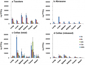
Fig 4. Comparison of biodistribution of free docetaxel (Taxotere), nab-PTX (Abraxane) and Cellax-DTX when infused 40mg taxane/kg. Cellax-DTX had preferable selective uptake and sustained concentrations for tumor compared to other formulations. From Yang et al. AAPS J 2017 Mar;19(2):386-396. Image adapted from Ernsting et al. Biomaterials 2012;33(5):1445-54 and Ernsting et al. JCR 2012;162(3):575-81.
Anti-tumor Efficacy
Cellax-DTX displays superior antitumor efficacy compared to free DTX and Nab-PTX in prostate cancer, breast cancer, lung cancer, pancreatic cancer and melanoma tumor models. (Yang et al., 2017: https://www.ncbi.nlm.nih.gov/pubmed/27873118)
Some of the findings have been shown below.
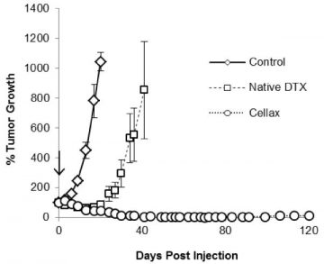
Fig 5. Cellax displays superior inhibition of tumor growth in a s.c. PC3 tumor xenograft model. Either Cellax (170mg DTX/kg), free DTX (20mg/kg) or saline was injected. Image from Hoang et al. Int J Pharm 2014;471(0):224-233. doi:10.1016/j.ijpharm.2014.05.021.
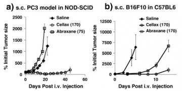
Fig 6. Cellax-DTX displays superior inhibition of tumor growth compared to Nab-PTX for prostate (PC3, a)) and melanoma(B16F10, b)) tumor models. In a), either a single i.v. dose of Abraxane 75mg PTX/kg was given or Cellax 170mg DTX/kg. In b), either single i.v. dose of Abraxane 170mg PTX/kg or Cellax 170mg DTX/kg was given. Image from Ernsting et al. JCR 2012;162(3):575-581. doi:10.1016/j.jconrel.2012.07.043.
Safety
Cellax-DTX displays a preferable safety profile compared to native DTX. The maximum tolerated dose (MTD) for Taxotere (native DTX) and Cellax-DTX was performed. In the NOD-SCID models, Taxotere had a MTD of 20mg/kg while Cellax had a MTD of 170mg/kg. Dose-limiting of native DTX includes neutropenia while the dose-limiting toxicity of Cellax is reversible liver inflammation. (Ernsting et al., 2012.: https://www.ncbi.nlm.nih.gov/pubmed/22369962)
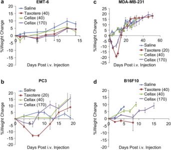
Fig 7. Cellax displayed milder, shorter weight loss than free docetaxel-treated mice. The dose of mg drug/kg is displayed in brackets. Image from Ernsting et al. Biomaterials 2012;33(15):3931-3941. doi:10.1016/j.biomaterials.2012.02.019.